Diagnosis
Saddle Embolus
Summary
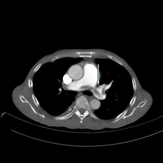
Axial image from a CT pulmonary angiogram demonstrates a saddle embolus at the bifurcation of the pulmonary trunk, extending into the right and left pulmonary arteries.
Saddle emboli are generally regarded as unstable emboli which may fragment and obstruct distal branches of the pulmonary arteries. Obstruction of more than 30% of the pulmonary circulation causes sufficient elevation of the pulmonary vascular resistance to produce significant pulmonary hypertension, resulting in increased afterload, right ventricle dilatation with decreased stroke volume, tricuspid regurgitation, reduced venous return and finally circulatory collapse.
1
2
There are radiologic findings associated with right heart strain, which may be evident.
Treatment options for a large pulmonary embolus include high dose unfractioned heparin, fibrinolysis and embolectomy.
3
References
-
Wood KE. Major pulmonary embolism: review of a pathophysiologic approach to the golden hour of hemodynamically significant pulmonary embolism. Chest 2002; 121: 877–905.
-
Ghaye B, Ghuysen A, Bruyere PJ, D’Orio V, Dondelinger RF. Can CT pulmonary angiography allow assessment of severity and prognosis in patients with pulmonary embolism?. Radiographics 2006; 26: 23-39.
-
Kucher N, Goldhaber SZ. Management of massive pulmonary embolus. Circulation. 2005; 112: e28-e32

