Diagnosis
Inactive Tuberculosis
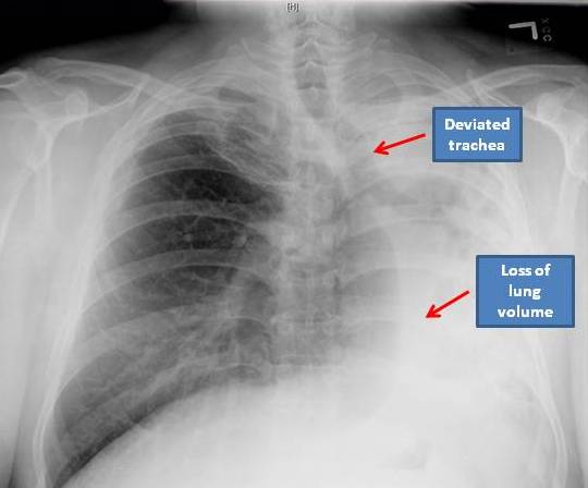
Summary
A 49 yr old Hispanic male moved to the Bronx, and came to the clinic for routine medical care. He has a history of hypertension, diabetes mellitus, and hyperlipidemia. Review of systems was only notable for an occasional dry cough. No history of weight loss, smoking or chemical exposure. Physical exam showed symmetric chest wall and clear to auscultation of the lung fields. PPD status unknown. HIV was nonreactive. Three samples of sputum are negative for Acid fast bacilli. Routine chest X ray (shown) had scarring and interstitial lung disease with loss of lung volume. The heart, mediastinum and trachea are deviated to the left. Bilateral Pleural disease is noted. Small rounded lucencies within the lateral aspect of the left chest noted. Past medical information revealed a past history of tuberculosis which was treated medically. Differential diagnosis for loss of lung volume in this patient includes TB, mediastinal lymphomas (obstruction of the bronchus secondary to tumors) and mucus plugging of the left lung. The diagnosis in this patient was consistent with inactive Tuberculosis. Chest X ray abnormalities suggestive of inactive TB are fibrotic scar with or without volume loss, calcified or non-calcified nodules, pleural thickening, blunt costo-phrenic angles, diaphragmatic tenting.