Radiology Case of the Month
November 2018
A 34-Year old male presents with acute onset headache, dizziness, nausea and vomiting for 4 days.
Quiz-summary
0 of 2 questions completed
Questions:
- 1
- 2
Information
You have already completed the quiz before. Hence you can not start it again.
Quiz is loading…
You must sign in or sign up to start the quiz.
You must first complete the following:
Results
Results
0 of 2 questions answered correctly
Time has elapsed
You have reached 0 of 0 point(s), (0)
Earned Point(s): 0 of 0, (0)
0 Essay(s) Pending (Possible Point(s): 0)
| Average score |
|
| Your score |
|
Categories
- Not categorized 0%
-

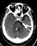
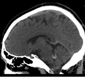
Noncontrast Head CT images demonstrate a focal area of hyperdensity about the medial aspect
of the right cerebellum adjacent to the fourth ventricle (yellow arrows).1.) With regards to the above images from non-contrast head CT, please select whichever applies:
→ A.) There is intracranial hemorrhage.
B.) There is a ruptured brain aneurysm
C.) There is acute obstructive hydrocephalus
D.) There is a meningioma of the roof the fourth ventricle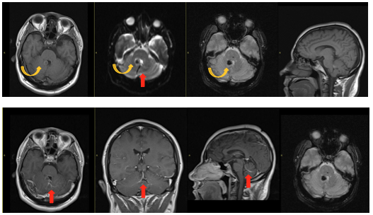
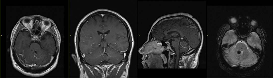
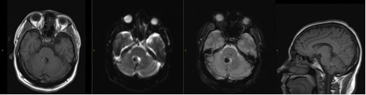
Postcontrast images demonstrate a focal area of blooming at the previously seen site of high density on CT at the right medial margin of the fourth ventricle without enhancement on postcontrast (yellow arrow) representing cavernoma. There is also demonstration of caput medusae appearance of small branching veins draining into a single vein adjacent to this lesion (red arrow) suggestive of a deep venous anomaly (DVA).
2.) With regards to the above images from MRI with and without contrast, please select whichever applies:
→ A.) There is a vascular malformation
B.) There is a thrombosed posterior circulation aneurysm
C.) There is an enhancing mass in the fourth ventricle
D.) This is a neurosurgical emergency and requires urgent endovascular coiling


- 1
- 2
- Answered
- Review
-
Question 1 of 2
1. Question

1.) With regards to the above images from non-contrast head CT, please select whichever applies:
CorrectIncorrect -
Question 2 of 2
2. Question

With regards to the above images from MRI with and without contrast, please select whichever apply:
CorrectIncorrect